Custom Search

How Does The Human Eye Work?

The individual components of the eye work in a manner similar to a camera. Each part plays a vital role in providing clear vision. So think of the eye as a camera with the cornea, behaving much like a lens cover. As the eye's main focusing element, the cornea takes widely diverging rays of light and bends them through the pupil, the dark, round opening in the center of the colored iris. The iris and pupil act like the aperture of a camera.

Next in line is the lens which acts like the lens in a camera, helping to focus light to the back of the eye. Note that the lens is the part which becomes cloudy and is removed during cataract surgery to be replaced by an artificial implant nowadays.

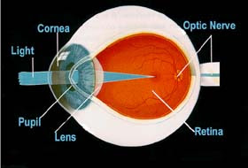


The very back of the eye is lined with a layer called the retina which acts very much like the film of the camera. The retina is a membrane containing photoreceptor nerve cells that lines the inside back wall of the eye. The photoreceptor nerve cells of the retina change the light rays into electrical impulses and send them through the optic nerve to the brain where an image is perceived. The center 10% of the retina is called the macula. This is responsible for
your sharp vision, your reading vision. The peripheral retina is responsible for the peripheral vision. As with the camera, if the "film" is bad in the eye (i.e. the retina), no matter how good the rest of the eye is, you will not get a good picture.

The human eye is remarkable. It accommodates to changing lighting conditions and focuses light rays originating from various distances from the eye. When all of the components of the eye function properly, light is converted to impulses and conveyed to the brain where an image is perceived.


Glossary of Eye Terms:


Anterior Chamber

The cavity in the front part of the eye between the lens and cornea is called the Anterior Chamber. It is filled with Aqueous, a water-like fluid. This fluid is produced by the ciliary body and drains back into the blood circulation through channels in the chamber angle. It is turned over every100 minutes.

Chamber Angle

Located at the junction of the cornea, iris, and sclera, the anterior chamber angle extends 360 degrees at the perimeter of the iris. Channels here allow aqueous fluid to drain back into the blood circulation from the eye. May be obstructed in glaucoma.

Ciliary Body

A structure located behind the iris (rarely visible) which produces aqueous fluid that fills the front part of the eye and thus maintains the eye pressure. It also allows focusing of the lens.

Conjunctiva

A thin lining over the sclera, or white part of the eye. This also lines the inside of the eyelids.Cell in the conjunctiva produce mucous, which helps to lubricate the eye.

Cornea

The transparent, outer "window" and primary focusing element of the eye. The outer layer of the cornea is known as epithelium. Its main job is to protect the eye. The epithelium is made up of transparent cells that have the ability to regenerate quickly. The inner layer of the cornea is also made up of transparent tissue, which allows light to pass.

Hyaloid Canal

A narrow channel that runs from the optic disc to the back surface of the lens. It serves an embryologic function prior to birth but none afterwards.

Iris

Inside the anterior chamber is the iris. This is the part of the eye which is responsible for one's eye color. It acts like the diaphragm of a camera, dilating and constricting the pupil to allow more or less light into the eye.

Pupil

The dark opening in the center of the colored iris that controls how much light enters the eye.The colored iris functions like the iris of a camera, opening and closing, to control the amount of light entering through the pupil.

Lens

The part of the eye immediately behind the iris that performs delicate focusing of light rays upon the retina. In persons under 40, the lens is soft and pliable, allowing for fine focusing from a wide variety of distances. For individuals over 40, the lens begins to become less pliable, making focusing upon objects near to the eye more difficult. This is known as presbyopia.

Macula

The part of the retina which is most sensitive, and is responsible for the central (or reading)vision. It is located near the optic nerve directly at the back of the eye (on the inside). This area is also responsible for color vision.

Optic Disc

The position in the back of the eye where the nerve (along with an artery and vein) enters the eye corresponds to the "blind spot" since there are no rods or cones in these location. Normally, a person does not notice this blind spot since rapid movements of the eye and processing in the brain compensate for this absent information. This is the area that the ophthalmologist studies when evaluating a patient for glaucoma, a condition where the optic nerve becomes damaged often due to high pressure within the eye. As it looks like a cupwhen viewed with an ophthalmoscope, it is sometimes referred to as the Optic Cup.

Optic Nerve

The optic nerve is the structure which takes the information from the retina as electrical signals and delivers it to the brain where this information is interpreted as a visual image. The optic nerve consists of a bundle of about one million nerve fibers.

Retina

The membrane lining the back of the eye that contains photoreceptor cells. These
photoreceptor nerve cells react to the presence and intensity of light by sending an impulse to the brain via the optic nerve. In the brain, the multitude of nerve impulses received from the photoreceptor cells in the retina are assimilated into an image.

Sclera

The white, tough wall of the eye. Few diseases affect this layer. It is covered by the episclera (a fibrous layer between the conjunctiva and sclera ) and conjunctiva, and eye muscles are connected to this.

Vitreous

Next in our voyage through the eye is the vitreous. This is a jelly-like substance that fills the body of the eye. It is normally clear. In early life, it is firmly attached to the retina behind it.With age, the vitreous becomes more water-like and may detach from the retina. Often, little clumps or strands of the jelly form and cast shadows which are perceived as "floaters". While frequently benign, sometimes floaters can be a sign of a more serious condition such as a retinal tear or detachment and should be investigated with a thorough ophthalmologic
examination.

Nutrition and the eye

Introduction

There has been a lot of interest in the role of nutrition and nutritional supplements, such as vitamin tablets, and their effect on vision. The main focus has been on how vitamins and minerals might affect age-related eye conditions such as age-related macular-degeneration (AMD) and cataracts.

This page explores these issues and looks at the importance of good nutrition and how the eye can be affected by it.

Importance of good nutrition

Good nutrition is very important for both your general and eye health. Good nutrition helps our body to grow, repair wear and tear, protect against infection and to function properly.

An eye condition called 'xerophthalmia', which is a common cause of childhood blindness in developing countries, is a good example of how nutrition and eye health go hand in hand. This condition is caused by a lack of vitamin A in a person's diet and could be prevented by eating fresh vegetables, fat (animal and plant) and protein (eg meat, eggs, cheese, fish, poultry, milk, yoghurt, dairy products, nuts, seeds, pulses and grains).

There are many books on nutrition and eating a balanced diet. This topic will only be looked at briefly here. For further information on nutrition and healthy eating contact the British Nutrition Foundation (BNF).

Nutrition for the eye

Vitamins and the eye.

In various studies and clinical trials antioxidant vitamins found in certain foods have been linked with eye health. They help to maintain healthy cells and tissues in the eye.

The main focus has been on the anti-oxidant vitamins A, C and E. These vitamins can be found in many different sources of fruit and vegetables such as:

•oranges
•kiwis
•grapefruit
•dried apricots
•tomatoes
•peppers
•raw carrots
•green leafy vegetables including kale and spinach
•green peas
•green beans
•brussel sprouts.
They can also be found in nuts, seeds, dairy products and eggs.

These are only a few of the food types in which antioxidant vitamins can be found. The British Nutrition Foundation can provide you with further information on this.

Lutein and eye health.

More recently it has been suggested that two types of antioxidants, known as 'carotenoids', called Lutein (pronounced Loo-teen) and Zeaxanthin (pronounced Zay-a-za-thin) may also help with eye health. Some studies have found that people who have a good diet rich in carotenoids, particularly lutein and zeaxanthin, have a lower risk of developing AMD.

Lutein and Zeaxanthin can be found naturally in vegetables and fruit. For example, Lutein can be found in yellow peppers, mango, bilberries, and green leafy vegetables such as kale, spinach, chard and broccoli.

Zeaxanthin can be found in orange sweet peppers, broccoli, corn, lettuce (not iceberg), spinach, tangerines, oranges and eggs.

Many of these overlap with food types in which vitamins A, E and C are present.

Vitamins supplements and diet

Age-related macular degeneration (AMD)

A large research trial, called the 'Age-Related Eye Disease Study' (AREDS), showed that high quantities of the antioxidant vitamins A, C, E, beta-carotene and the minerals zinc as zinc oxide, and copper as cupric oxide, can help to slow down the progression of AMD.

It would be very hard to obtain the large quantity of vitamins used in the trial from your diet. Therefore some people who have AMD may consider supplementation with vitamins and anti-oxidants. Such high dosages of vitamins and minerals might have possible side effects on the body.

For this reason it is very important to consult your doctor first before taking a supplement.

Eye health

Following the AREDS research trial there have been over 150 smaller scale studies looking at how vitamins and minerals, both from food and in a vitamin supplement, can help eye health in general, and in particular AMD and cataracts. A number of these studies have looked specifically at the carotenoids Lutein and Zeaxanthin which have been particularly associated with healthy eyes.

Some of these studies have shown how certain vitamin and mineral supplements can have a positive effect on eyes and sight. Others have shown there to be no or little benefit. For this reason various organisations are calling for further, larger scale research.

As a result of these studies there are now a number of different supplements for eye health on the market. There is still divided medical opinion on the use of supplements for both eye health and for preventing, or slowing down, the progression of AMD and cataracts in particular.

The general consensus of opinion is that with a good balanced diet that includes sufficient fresh fruits and vegetables there should be no need to use supplements.

However, research has shown that many people in the UK do not get enough vitamins and minerals from their diet. Some people might consider taking a supplement for their general and eye health when:

•their diet does not include enough fresh fruit and vegetables
•diet does not include enough vitamins and minerals
•vitamins and minerals from food are not adequately absorbed by the body
•it is hard to obtain or prepare fresh fruit and vegetables
•they have been told to take a supplement by their doctor or nutritionist.
However, experts agree that taking supplements is not a substitute for a healthy diet.

Conclusion

Evidence regarding the benefits of nutritional supplements against eye disease is conflicting and there is no real agreement among researchers on this subject at present.

However, a consensus has been reached on the importance of a healthy, balanced diet full of fresh fruits and vegetables, particularly leafy green vegetables such as kale and spinach.

Key points to remember:

•eat a good, balanced diet with lots of fresh fruit and vegetables
•discuss changing your diet or taking vitamin supplements with your GP
•discuss your diet or taking a vitamin supplement with your GP if you believe that your diet may be inadequate
•the biggest avoidable risk is smoking
•protect your eyes from sunlight. Use good quality sunglasses, ie those that have the 'CE' mark, which means they meet the European Union Quality Standards. Wearing a brimmed hat also offers very good protection
•get your eyes tested at least every two years and more frequently if necessary.

Eye Problems & Children

Eye problems are common in children, and range from infections, irritation, allergies to changes in vision. Any injury to the eye or problem that is affecting your child's vision should be evaluated by a doctor right away.

Conjunctivitis: conjunctivitis or pinkeye, is an infection caused by a virus or bacteria, that can cause redness in one or both eyes and a watery discharge. Bacterial conjunctivitis causes a red eye and matting of the eyes with a green or yellow discharge and it requires treatment with antibiotics (usually a topical ointment or drops). Conjunctivitis caused by a virus can also produce a red eye, but the discharge is usually just watery or cloudy. This type of infection is usually accompanied by symptoms of an upper respiratory tract infection, such as a runny nose and cough, and he may have an enlarged lymph gland in front of his ear. No treatment is usually required for this type of infection.
Allergic Conjunctivitis: conjunctivitis can also be caused by allergies, especially in children with allergies or asthma, and can cause both eyes to be red and swollen, itching and tearing. Antihistamines or eye drops may be needed for treatment.

Stye: A stye or hordeolum produces a tender, red swollen area on your child's eyelid. It may point inward or it may be on the outside of the eyelid margin. Treatment is with warm compresses and sometimes topical antibiotics.

Chalazion: If there is a nodule or swollen area on the eyelid, but it isn't red or tender, then it is probably a chalazion, which is an inflamed meibomian gland that improves by using warm compresses several times each day for several weeks or months. Persistent cases sometimes require excision.
Corneal Abrasion: can occur after having a foreign body in the eye or after scratching the eye. This causes pain, tearing, and sensitivity to light. An abrasion can be detected by your pediatrician by putting fluorescein dye drops in the eye and looking at it with a special blue light. Treatment is with antibiotic ointment until the abrasion has healed, unless the abrasion is over the pupil, in which case your child should probably see an ophthalmologist.

Blepharitis: an inflammation of the eyelids that causes them to have redness, crusting and scaling and the eyes may be itchy or irritated. Treatment is usually with daily cleansing of the eyelids with a no-tears shampoo and a soft cloth. Sometimes an antibiotic cream is also needed. Untreated, this can lead to the formation of a stye or chalazion.

Foreign body: having a foreign object in the eye, such as a piece of sand, etc, can cause pain,, especially with blinking, and tearing. Older children may complain that it feels like there is something in the eye. Treatment is by washing the object out of the eye with lukewarm water. See you pediatrician if that doesn't help.

Dacryostenosis: This is a blocked tear duct, are very common in newborns and infants and usually clear up on their own by the time your baby is a year old. It can cause some matting of the eye, and persistent tearing in one or both eyes. You should wipe away any discharge with a warm cloth. You can also gently massage the side of the upper part of your baby's nose to try and open up the duct. If your baby has a lot of discharge that needs to be wiped away more than a few times a day, then he may need an antibiotic and you should see your doctor.
Subconjunctival hemorrhages: occur because of pressure during the birth process. It causes a red streaking or red spots of blood on the white part of the eye and it will go away in a few weeks. It can also occur in older children after vomiting forcibly.

Periorbital cellulitis: This is a serious infection that can cause a fever and your child's eyelids to be red, swollen and tender. Left untreated, it can progress to orbital cellulitis or a deep infection of the eye, which can cause the eye to be pushed outwards (proptosis), and may limit the movement of the eye. You should seek medical attention immediately for either of these conditions.
Other eye problems include those caused by trauma, including fractures to the bones around the eye, chemical burns, and lacerations or cuts to the eye or eyelids. These require immediate medical attention.

Parts of the eye


Basic functions of the human eye

The basic function of the human eye is to see. Other functions involving the accessory structures and secretions of the eye prevent foreign particles from injuring the ocular organ and pathogens from entering the body through this route.

In its simplest definition, sight is the transmission of light signals through nerve endings to the brain, which interprets the signals and produces visions of what is seen. The light reaches the nerve endings by traveling through the eye, making the brain's interpretation dependent on the accuracy of the path the light waves travel through the structures of the eye.

Human TissueHuge Selection. Great value 4 less! Many Species, 5000+ Tissue Types.www.BioChain.comHuman BrainAnswers to your questions on Alzheimer's Disease and more.yourtotalhealth.ivillage.comAdvanced BioMatrix, Inc.PureCol Purified Collagen Products 3D Matrix Cell Culture Reagentswww.advancedbiomatrix.comLight's

Path Through the Eye

Light enters the eye first through the cornea, a thin clear sheath covering the outer, visible portion of the eye. There are no blood vessels in the cornea because it must remain clear for light waves to pass through uninterrupted. Under the cornea is a cavity containing a nutrient-filled liquid, aqueous humor, which provides nutrients to the cornea and lens. The pupil is the opening that controls how much light passes from the cornea to the lens. The iris, the colored portion of the eye, is actually two sets of muscle fibers that control the size of the pupil.

The lens is the refraction system of the eye; it is located on the other side of the aqueous humor cavity. The lens also contains no blood vessels so that light can pass through unhindered. Light is refracted and focused onto the back of the eye where the nerve endings are present; what is seen may be out of focus if the lens is misshapen or the wrong thickness, resulting in the vision disorders near-sightedness, far-sightedness, and stigmatism. The lens is adjusted by muscles called the ciliary body, which allow light to be focused for distance and movement.

Light travels from the lens through a chamber filled with vitreous humor, a semi-solid that helps the eye keep its shape. The vitreous humor is also what holds the retina in place. The retina is the back of the inside of the eye and contains light receptors. The receptors are nerve endings with a direct link to the brain via the optic nerve. Alterations in the vitreal pressure due to eye injuries or diabetes can affect vision and cause retinal detachment or blindness.

Light Receptors How Vision Works

Receptors in the eye consist of rods and cones. Rods detect light regardless of wavelength and help the brain determine light intensity, whether it is bright or dim. Cones are only active in bright light and are particular to red, blue, or green. Each type of cone detects a particular light wavelength. Color blindness is a disorder that occurs when a person lacks a particular type or types of cones, allowing only bright, dim, and the remaining wavelengths to be seen.

There is a blind spot at the back of the eye at the point where the optic nerve passes through the eyeball and there are no receptors. With two working eyes, the blind spot is compensated for by binocular vision. There is also a spot in the retina with the greatest concentration of cones, called the macula fovea. This specialized pit in the 5-mm wide macula is important for high visual acuity. The macula has two layers of ganglion cells, the nerve endings communicating with the brain via the optic nerve.

Other Functions of the Eye

The eye has accessory structures. The eyelids have hairs called eyelashes that protect the eye and keep out dust and harmful debris. Scratching the cornea would prevent light from passing through properly and hinder vision. Tear ducts, called lacrimal glands, secrete a salt solution to keep the eye from drying out as well as a lysozyme that protects the eye from bacteria

Common Eye Problems

Most of us will experience temporary eye problems from time to time, including itching, blurriness or fatigue. Most of these conditions are short-lived and will probably go away on their own with no complications. However, sudden eye problems and those that last for more than a couple of days should be checked by an eye doctor. The following is a list of common eye problems and their possible causes.

Eye twitching:

While usually harmless, an eye twitch can be very annoying. Twitching may be associated with the following:

•Stress
•Caffeine
•Fatigue
•Pink eye
•Panic disorder
•Tourette syndrome

Itchy eyes:

Are your eyes itching? Itchy, red eyes can be very uncomfortable. Here are a few conditions that may cause itchy eyes:

•Pink eye
•Blepharitis
•Dry eye syndrome
•Allergies

Tired eyes:

Do your eyes feel tired and fatigued? The following conditions may cause tired eyes:

•Dry eye syndrome
•Computer vision syndrome
•Farsightedness
•Astigmatism

Seeing floaters:

Do you see spots in your vision? Do you see faint strands floating around that seem to move away if you look at them? Floaters may occur with the following conditions:

•Retinal detachment
•Diabetic retinopathy
•Posterior vitreous detachment
•Stroke

Blurry vision:

Blurry vision, or the inability to bring objects into sharp focus, can be caused by a number of conditions. Sometimes fatigue or illness can cause vision to be temporarily blurred. The following eye problems can also cause blurry vision:

•Nearsightedness
•Farsightedness
•Presbyopia
•Astigmatism
•Cataracts
•Glaucoma
•Uveitis
•Dry eye syndrome
•Migraine

Age-related problems:

Many eye problems occur as we get older. As always, annual comprehensive eye examinations are important for maintaining healthy vision as we age. The following eye problems may develop due to aging:

•Age-related macular degeneration
•Presbyopia
•Cataracts
•Glaucoma
•Posterior vitreous detachment

Children's eye problems:

Parents are often the first to notice eye problems in their children. Alert an eye care professional if you notice possible problems with your child's eyes or vision. The following eye problems may be responsible:

•Lazy eye
•Strabismus
•Color blindness
•Pink eye
•Farsightedness

Eye Health Is Important Throughout Our Lives

Keeping our eyes healthy is important throughout our lives. Learn how to protect your vision, prevent and treat eye diseases, and prevent eye injuries. Find out all about the different eye care professionals. Along with eye examinations, there are many things you can do to keep your eyes healthy.

Protecting Your Vision
Eye Injuries
Eye Diseases
Eye Problems